Bursitis After Foot Surgery
Overview
Whenever tissues rub against one another, a bursa forms to allow for smooth gliding. A bursa is a fluid-filled sac and occurs normally throughout the body. Only a few cell layers thick, a bursa is filled with a lubricating fluid. However, when irritated, a bursa can become markedly thickened and painful. This is often referred to as bursitis. The retrocalcaneal bursa is positioned to allow the Achilles tendon to glide over the back part (posterior aspect) of the heel bone. When this bone becomes enlarged, inflammation of the retrocalcaneal bursa occurs. This inflammation results in exquisite tenderness along the posterior aspect of the heel.
Causes
Feet are extremely resilient and are designed to stand up to the pressures of day-to-day living. In some cases, though, foot structures may break down when subjected to chronic stress associated with long periods of weight-bearing activity on concrete, asphalt, or other hard surfaces (especially when footwear does not allow for proper weight distribution). Foot problems, including infracalcaneal bursitis, are often made worse by poorly designed footwear, and pressure, impact, and shear forces can damage the feet over time. Bursal sacs are intended to minimize this damage, but sometimes the bursa itself becomes inflamed. A rapid increase in physical activity levels or thinning of the heel?s protective fat pad are factors that may contribute to infracalcaneal bursitis. Other possible causes of infracalcaneal bursitis include blunt force trauma. Arthritic conditions. Acute or chronic infection. The following factors may increase a person?s risk of bursitis, including infracalcaneal bursitis. Poor conditioning. Exposure to cold weather. Participating in contact sports. Having a previous history of bursitis in any joint. Heel striking when running, especially in conventional running shoes with heel elevation.
Symptoms
Pain or tenderness at the back of the heel around the Achilles region. Increased pain during activities with strong, repetitive calf contractions, walking (uphill), stair climbing, running, jumping. Pain may be worse with rest after activity (that night or the next morning) or at the beginning of the excercise. Pain when wearing shoes and the heel is getting rubbed. Bump forming on the back of the heel. Limping. Stiffness. Decreased range of motion. Redness and warmth (if the bursa gets infected).
Diagnosis
The diagnosis is based on the symptoms and an examination. For anterior Achilles tendon bursitis, doctors use x-rays to rule out a fracture of the heel bone or damage to the heel bone caused by rheumatoid arthritis or other inflammatory arthritis.
Non Surgical Treatment
Many times, Achilles tendon bursitis can be treated with home care. However, if self-care remedies do not work, your doctor may need to administer additional treatments. Home care. The most important thing you can do to help your healing is to refrain from activities that could further aggravate the bursitis. Typical home-care treatments for Achilles tendon bursitis include Anti-inflammatory medication. Take over-the-counter pain medication, like aspirin or ibuprofen, to reduce pain and swelling. Heel wedges. An over-the-counter or custom heel wedge can be placed in your shoe to minimize stress in the Achilles tendon. Ice. Apply ice or other cold therapy to your sore heel several times a day. Rest. Limit your activity on the injured leg, avoid putting pressure on it whenever possible. Also, elevate your leg during non-use to help reduce the swelling. The above remedies may be used on their own or in combination with others on the list. Physician-administered treatments. If the above self-care remedies are not effective, you should visit your doctor for additional treatment. Possible Achilles tendon bursitis treatments your doctor may administer include immobilization. If the bursitis is combined with Achilles tendonitis, your doctor may place a temporary cast on the ankle to prevent movement and allow it to heal. Physical therapy. Exercises may be used to improve the ankle's strength and flexibility. Steroids. Injection of steroids into the retrocalcaneal bursa (not the Achilles tendon) may be necessary. Surgery. In very rare circumstances, surgery may be needed to remove the retrocalcaneal bursa, however, this is typically a last resort.
Surgical Treatment
Bursectomy is a surgical procedure used to remove an inflamed or infected bursa, which is a fluid-filled sac that reduces friction between tissues of the body. Because retrocalcaneal bursitis can cause chronic inflammation, pain and discomfort, bursectomy may be used as a treatment for the condition when it is persistent and cannot be relived with other treatments. During this procedure, a surgeon makes small incisions so that a camera may be inserted into the joint. This camera is called an arthroscope. Another small incision is made so that surgical instruments can be inserted to remove the inflamed bursa.
Hammer Toe Fusion
 Overview
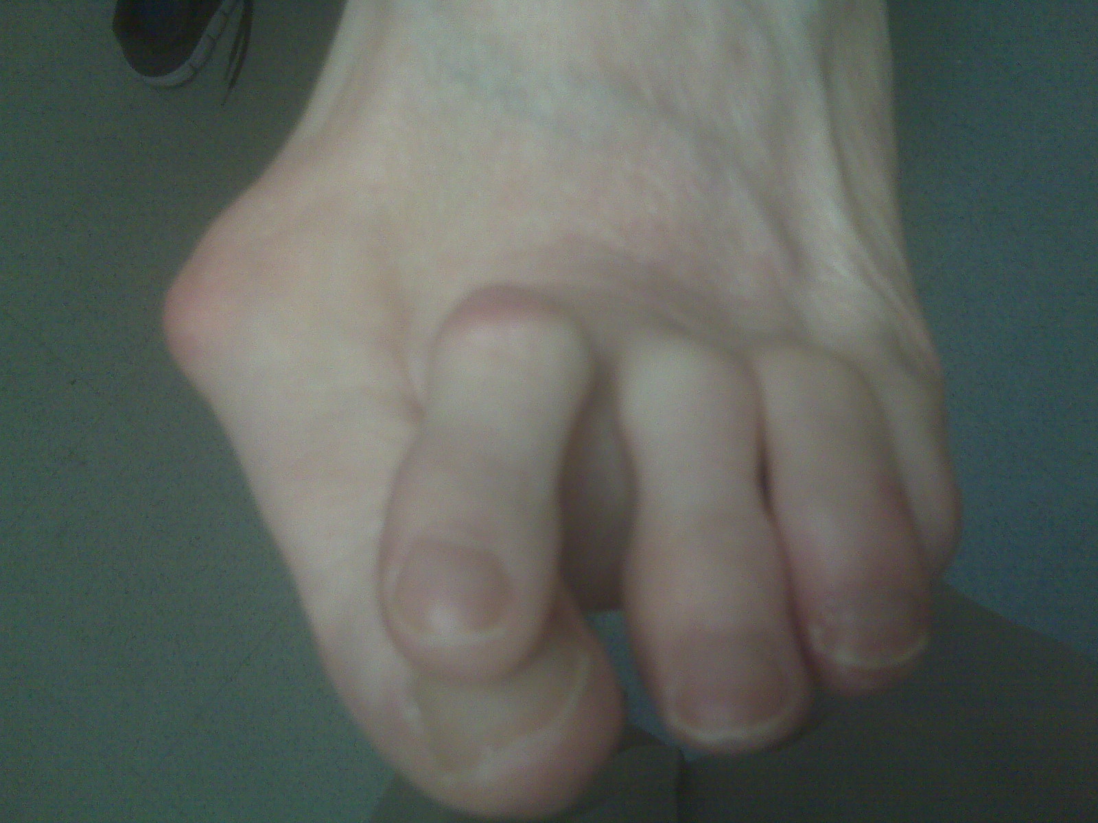
Hammer toe, also called hammer toe, deformity of the second, third, or fourth toe in which the toe is bent downward at the middle joint (the proximal interphalangeal [PIP] joint), such that the overall shape of the toe resembles a hammer. Most cases of hammertoe involve the second toe, and often only one or two toes are affected. In rare cases when all the toes are involved, a thorough neurological assessment is necessary to evaluate for underlying nerve or spinal cord problems.
Causes
Medical problems, such as stroke or diabetes that affect the nerves, may also lead to hammertoe. For example, diabetes can result in poor circulation, especially in the feet. As a result, the person may not feel that their toes are bent into unnatural positions. The likelihood of developing hammertoe increases with age and may be affected by gender (more common in women) and toe length; for example, when the second toe is longer than the big toe, hammertoe is more likely to occur. Hammertoe may also be present at birth. Genetics may factor in to developing hammertoe, particularly if the foot is flat or has a high arch, resulting in instability.
 Symptoms
Common symptoms of hammertoes include pain or irritation of the affected toe when wearing shoes. corns and calluses (a buildup of skin) on the toe, between two toes, or on the ball of the foot. Corns are caused by constant friction against the shoe. They may be soft or hard, depending upon their location. Inflammation, redness, or a burning sensation. Contracture of the toe. In more severe cases of hammertoe, open sores may form.
Diagnosis
Most health care professionals can diagnose hammertoe simply by examining your toes and feet. X-rays of the feet are not needed to diagnose hammertoe, but they may be useful to look for signs of some types of arthritis (such as rheumatoid arthritis) or other disorders that can cause hammertoe. If the deformed toe is very painful, your doctor may recommend that you have a fluid sample withdrawn from the joint with a needle so the fluid can be checked for signs of infection or gout (arthritis from crystal deposits).
Non Surgical Treatment
Symptomatic treatment of hammertoes consists of such things as open toed shoes or hammertoe pads. There are over the counter corn removers for temporally reducing the painful callous often seen with the hammertoe. These medications must be used with caution. They are a mild acid that burns the callous off. These medications should Hammer toe never be used for corns or callouses between the toes. Persons with diabetes or bad circulation should never use these products.
Surgical Treatment
If conservative treatments don't help, your doctor may recommend surgery to release the tendon that's preventing your toe from lying flat. In some cases, your doctor might also remove some pieces of bone to straighten your toe.
Causes Of Bunion Pain?
Overview
 A bunion is a very common foot deformity that develops over the first metatarsal phalageal joint of the big toe of the foot. The joint that joins the big toe to the foot is called the first MTP joint. When it becomes prominent and the big toe starts to become crooked this is known as a bunion. The term referring to deformity of the big toe as it becomes crooked is called Hallux Valgus. It is the bump itself that is known as the bunion. When it gets red and swollen over the bunion because it gets sore this is usually due to an inflamed soft tissue over the underlying bone.
Causes
The most common cause of bunions is wearing shoes that are too tight. The condition is not hereditary, but it does tend to run in families-often due to a faulty foot structure. Other causes of this condition might be neuromuscular problems, flat feet, pronated feet, or foot injuries.
Symptoms
A bulging bump on the outside of the base of your big toe. Swelling, redness or soreness around your big toe joint aggravated by footwear. Red, calloused skin along the inside edge of the big toe. Corns or calluses under the ball of the foot or where the first and second toes overlap. Persistent or intermittent pain. Restricted movement of your big toe.
Diagnosis
Generally, observation is adequate to diagnose a bunion, as the bump is obvious on the side of the foot or base of the big toe. However, your physician may order X-rays that will show the extent of the deformity of the foot.
Non Surgical Treatment
Wearing good footwear does not cure the deformity but may ease symptoms of pain and discomfort. Ideally, get footwear advice from a person qualified to diagnose and treat foot disorders (podiatrist - previously called a chiropodist). Advice may include wear shoes, trainers or slippers that fit well and are roomy. Don't wear high-heeled, pointed or tight shoes. You might find that shoes with laces or straps are best, as they can be adjusted to the width of your foot. Padding over the bunion may help, as may ice packs. Devices which help to straighten the toe (orthoses) are still occasionally recommended, although trials investigating their use have not found them much better than no treatment at all. Painkillers such as paracetamol or ibuprofen may ease any pain. If the bunion (hallux valgus) develops as part of an arthritis then other medication may be advised. A course of antibiotics may be needed if the skin and tissues over the deformity become infected. 
Surgical Treatment
The choice of surgical procedures (bunionectomy) is based on a biomechanical and radiographic examination of the foot. Because there is actual bone displacement and joint adaptation, most successful bunionectomies require cutting and realigning the 1st metatarsal (an osteotomy). Simply "shaving the bump" is often inadequate in providing long-term relief of symptoms and in some cases can actually cause the bunion to progress faster. The most common procedure performed for the correction of bunions is the 1st metatarsal neck osteotomy, near the level of the joint. This refers to the anatomical site on the 1st metatarsal where the actual bone cut is made. Other procedures are preformed in the shaft of the metatarsal bone (see procedures preformed in the shaft of the metatarsal) and still other procedures are selected by the surgeon that are preformed in the base of the metatarsal bone (see surgeries preformed in the base of the metatarsal).
Prevention
To help prevent bunions, select your style and size of shoes wisely. Choose shoes with a wide toe area and a half-inch of space between the tip of your longest toe and the end of the shoe. Shoes also should conform to the shape of your feet without causing too much pressure.
Over-Pronation Of The Foot What Are The Symptoms
Overview
Overpronation is when the foot rolls in excessively, or at a time when it should not, for instance late in the stance phase of gait. In this case much weight is transferred to the inner or medial side of the foot, and as the runner moves forward the load is borne by the inner edge rather than the ball of the foot. This destabilises the foot, which will attempt to regain stability by compensating for the inward movement. In a kind of chain reaction, this in turn affects the biomechanical efficiency of the leg, especially the knee and hip.
Causes
Although there are many factors that can contribute to the development of these conditions, improper biomechanics of the body plays a large and detrimental role in the process. Of the many biomechanical elements involved, foot and ankle function perhaps contribute the most to these aches and pains.
Symptoms
People with overpronation may suffer from pain in the knees, hips, and low back. Overpronation itself does not necessarily cause any other symptoms but is a contributing factor of many foot conditions such as Plantar Facsiitis, Heel Spur Syndrome, Posterior Tibialis Tendon Rupture or Tendonitis, Hallux Valgus, Bunion Deformities, Metatarsalgia, Hallux Limitus or Hallux Rigidus, Hammer Toes, and Morton?s Neuroma.
Diagnosis
A quick way to see if you over-pronate is to look for these signs. While standing straight with bare feet on the floor, look so see if the inside of your arch or sole touches the floor. Take a look at your hiking or running shoes; look for wear on the inside of the sole. Wet your feet and walk on a surface that will show the foot mark. If you have a neutral foot you should see your heel connected to the ball of your foot by a mark roughly half of width of your sole. If you over-pronate you will see greater than half and up to the full width of your sole.
Non Surgical Treatment
Solutions typically presented will include physical therapy sessions, prolonged prescription drug regimens, occasionally non-traditional approaches like holistic medicine and acupuncture. These options can provide symptom relief in the short term for some patients. However, these treatment methods cannot correct the internal osseous misalignment. Ligaments are not effective in limiting the motion of the ankle bone when excessive joint motion is present. Furthermore, there is not a single, specific ligament that is "too tight" that needs to be "stretched out." The muscles supporting the bones are already being "over-worked" and they cannot be strengthened enough to realign these bones. There is no evidence to suggest that any of these measures are effective in re-establishing or maintaining the normal joint alignment and function.
Prevention
Duck stance: Stand with your heels together and feet turned out. Tighten the buttock muscles, slightly tilt your pelvis forwards and try to rotate your legs outwards. You should feel your arches rising while you do this exercise.
Calf stretch:Stand facing a wall and place hands on it for support. Lean forwards until stretch is felt in the calves. Hold for 30 seconds. Bend at knees and hold for a further 30 seconds. Repeat 5 times.
Golf ball:While drawing your toes upwards towards your shins, roll a golf ball under the foot between 30 and 60 seconds. If you find a painful point, keep rolling the ball on that spot for 10 seconds.
Big toe push: Stand with your ankles in a neutral position (without rolling the foot inwards). Push down with your big toe but do not let the ankle roll inwards or the arch collapse. Hold for 5 seconds. Repeat 10 times. Build up to longer times and fewer repetitions.
Ankle strengthener: Place a ball between your foot and a wall. Sitting down and keeping your toes pointed upwards, press the outside of the foot against the ball, as though pushing it into the wall. Hold for 5 seconds and repeat 10 times.
Arch strengthener: Stand on one foot on the floor. The movements needed to remain balanced will strengthen the arch. When you are able to balance for 30 seconds, start doing this exercise using a wobble board.
Severs Disease Rehab
Overview
Pain in the heel of a child's foot, typically brought on by some form of injury or trauma, is sometimes Sever's Disease. The disease often mimics Achilles tendonitis, an inflammation of the tendon attached to the back of the heel. A tight Achilles tendon may contribute to Sever's Disease by pulling excessively on the growth plate of the heel bone. This condition is most common in younger children and is frequently seen in the active soccer, football or baseball player. Sport shoes with cleats are also known to aggravate the condition. Treatment includes calf muscle stretching exercises, heel cushions in the shoes, and/or anti-inflammatory medications. Consult your physician before taking any medications.
Causes
This condition is more common in boys than girls. It generally presents between the ages of 9-14 and peaks between ages 10-12 years. This injury can reoccur up until the age of 17, when the growth plate of the calcaneous generally closes. These types of injuries will commonly occur during periods of rapid growth. Sever?s Disease occurs more frequently in child with flat feet, but all children with flat feet will not get Sever?s.
Symptoms
The most obvious sign of Sever's disease is pain or tenderness in one or both heels, usually at the back. The pain also might extend to the sides and bottom of the heel, ending near the arch of the foot. A child also may have these related problems, swelling and redness in the heel, difficulty walking, discomfort or stiffness in the feet upon awaking, discomfort when the heel is squeezed on both sides, an unusual walk, such as walking with a limp or on tiptoes to avoid putting pressure on the heel. Symptoms are usually worse during or after activity and get better with rest.
Diagnosis
A Podiatrist can easily evaluate your child?s foot, lower limbs and muscular flexibility, to identify if a problem exists. If a problem is identified, a simple treatment plan is put in place. Initial treatment may involve using temporary padding and strapping to control motion or to cushion the painful area and based on the success of this treatment, a long-term treatment plan will be put in place. This long-term treatment plan may or may not involve Foot Supports, Heel Raises, muscle stretching and or strengthening.
Non Surgical Treatment
The aims are to reduce trauma to the heel, allow rest/recovery and prevent recurrence. Most cases are successfully treated using physiotherapy and exercises, eg to stretch the gastrocnemius-soleus complex, to mobilise the ankle mortise, subtalar joint and medial forefoot. Soft orthotics or heel cups. Advice on suitable athletic footwear. Other modes of treatment are in severe cases, temporarily limiting activity such as running and jumping. Ice and non-steroidal anti-inflammatory drugs (NSAIDs), which can reduce pain. In very severe cases, a short period of immobilisation (eg 2-3 weeks in a case in mild equinus position) has been suggested.
Exercise
For children with Sever's disease, it is important to habitually perform exercises to stretch the hamstrings, calf muscles, and the tendons on the back of the leg. Stretching should be performed 2-3 times a day. Each stretch should be performed for 20 seconds, and both legs should be stretched, even if the pain is only in one heel. Heel cups or an inner shoe heel lifts are often recommended for patient suffering from Sever's disease. Wearing running shoes with built in heel cups can also decrease the symptoms because they can help soften the impact on the heel when walking, running, or standing.
Arch Support Pain Relief
Overview
Pain across the bottom of the foot at any point between the heel and the ball of the foot is often referred to as "arch pain." Although this description is non-specific, most arch pain is due to strain or inflammation of the plantar fascia (a long ligament on the bottom of the foot). This condition is known as plantar fasciitis and is sometimes associated with a heel spur. In most cases, arch pain develops from overuse, unsupportive shoes, weight gain, or acute injury. If arch pain persists beyond a few days, see a foot and ankle surgeon for treatment to prevent this condition from becoming worse.

Causes
Spending a lot of time on your feet. Especially when you are not used to doing so. For example you may have started a new job such as waiting tables where you are on your feet all day and wake up the next day with sore feet. This is a sign of damage and over time could lead to plantar fasciitis. Being Over-Weight. Never an easy topic to discuss but in simple terms, the heavier you are, the greater the burden on your feet. There are times when you're walking when your entire body weight is borne on one leg and therefore one foot, placing great strain on the plantar fascia. Wearing shoes with poor arch support or cushioning. A tight Achilles tendon. This is the big tendon at the bottom of your calf muscles above your heel. If this is excessively tight this can affect your ability to flex your ankle and make you more likely to damage your plantar fascia. Suddenly changing your exercise routine. Using running as an example if you suddenly run many more miles than your are used to or change to a new running surface e.g. grass to tarmac, these factors can put excessive strain on the plantar fascia and lead to plantar fasciitis. All of these risk factors ultimately lead to a specific change in foot structure. The term given is over-pronation and this basically describes rolling in of the foot and lowering of the arches. It is this change that excessively elongates the plantar fascia which can lead to plantar fasciitis.
Symptoms
Many people have no symptoms, and the condition is discovered only by chance when an X-ray of the foot is obtained for some other problem. When symptoms occur, there is usually foot pain that begins at the outside rear of the foot. The pain tends to spread upward to the outer ankle and to the outside portion of the lower leg. Symptoms usually start during a child's teenage years and are aggravated by playing sports or walking on uneven ground. In some cases, the condition is discovered when a child is evaluated for unusually frequent ankle sprains.
Diagnosis
The doctor will take a brief history to determine how the injury occurred. If necessary, a thorough physical exam may be conducted to evaluate for any other injuries. Taking your workout shoes to the exam may also provide valuable information to the medical practitioner. Both feet will be physically and visually examined by the medical practitioner. The foot and arch will be touched and manipulated possibly with a lot of pressure and inspected to identify obvious deformities, tender spots, or any differences in the bones of the foot and arch.
Non Surgical Treatment
Treatment for a high arch foot or Charcot Marie Tooth disorder depends on the extent of deformity and the amount of disability experienced by the patient. Depending upon the symptoms, treatment may include. Changing the shoes. Special orthotic supports (devices that support, adjust, or accommodate the foot deformity). Cushioning pads. Foot and ankle braces or surgery.

Surgical Treatment
If pain or foot damage is severe, your doctor may recommend surgery. Procedures may include the following. Fusing foot or ankle bones together (arthrodesis). Removing bones or bony growths also called spurs (excision). Cutting or changing the shape of the bone (osteotomy). Cleaning the tendons' protective coverings (synovectomy). Adding tendon from other parts of your body to tendons in your foot to help balance the "pull" of the tendons and form an arch (tendon transfer). Grafting bone to your foot to make the arch rise more naturally (lateral column lengthening).
Stretching Exercises
People with flexible feet who develop fallen arches may benefit from foot strengthening exercises, notes the Nicholas Institute of Sports Medicine and Athletic Trauma. Standing on a towel in bare feet and grasping the material with the toes is an easy foot-strengthening exercise that can be done at home. Standing on one leg while arching and releasing the foot may also prove useful. Doctors may prescribe gentle stretching exercises for the foot and ankle tendons.
Pain across the bottom of the foot at any point between the heel and the ball of the foot is often referred to as "arch pain." Although this description is non-specific, most arch pain is due to strain or inflammation of the plantar fascia (a long ligament on the bottom of the foot). This condition is known as plantar fasciitis and is sometimes associated with a heel spur. In most cases, arch pain develops from overuse, unsupportive shoes, weight gain, or acute injury. If arch pain persists beyond a few days, see a foot and ankle surgeon for treatment to prevent this condition from becoming worse.
Causes
Spending a lot of time on your feet. Especially when you are not used to doing so. For example you may have started a new job such as waiting tables where you are on your feet all day and wake up the next day with sore feet. This is a sign of damage and over time could lead to plantar fasciitis. Being Over-Weight. Never an easy topic to discuss but in simple terms, the heavier you are, the greater the burden on your feet. There are times when you're walking when your entire body weight is borne on one leg and therefore one foot, placing great strain on the plantar fascia. Wearing shoes with poor arch support or cushioning. A tight Achilles tendon. This is the big tendon at the bottom of your calf muscles above your heel. If this is excessively tight this can affect your ability to flex your ankle and make you more likely to damage your plantar fascia. Suddenly changing your exercise routine. Using running as an example if you suddenly run many more miles than your are used to or change to a new running surface e.g. grass to tarmac, these factors can put excessive strain on the plantar fascia and lead to plantar fasciitis. All of these risk factors ultimately lead to a specific change in foot structure. The term given is over-pronation and this basically describes rolling in of the foot and lowering of the arches. It is this change that excessively elongates the plantar fascia which can lead to plantar fasciitis.
Symptoms
Many people have no symptoms, and the condition is discovered only by chance when an X-ray of the foot is obtained for some other problem. When symptoms occur, there is usually foot pain that begins at the outside rear of the foot. The pain tends to spread upward to the outer ankle and to the outside portion of the lower leg. Symptoms usually start during a child's teenage years and are aggravated by playing sports or walking on uneven ground. In some cases, the condition is discovered when a child is evaluated for unusually frequent ankle sprains.
Diagnosis
The doctor will take a brief history to determine how the injury occurred. If necessary, a thorough physical exam may be conducted to evaluate for any other injuries. Taking your workout shoes to the exam may also provide valuable information to the medical practitioner. Both feet will be physically and visually examined by the medical practitioner. The foot and arch will be touched and manipulated possibly with a lot of pressure and inspected to identify obvious deformities, tender spots, or any differences in the bones of the foot and arch.
Non Surgical Treatment
Treatment for a high arch foot or Charcot Marie Tooth disorder depends on the extent of deformity and the amount of disability experienced by the patient. Depending upon the symptoms, treatment may include. Changing the shoes. Special orthotic supports (devices that support, adjust, or accommodate the foot deformity). Cushioning pads. Foot and ankle braces or surgery.
Surgical Treatment
If pain or foot damage is severe, your doctor may recommend surgery. Procedures may include the following. Fusing foot or ankle bones together (arthrodesis). Removing bones or bony growths also called spurs (excision). Cutting or changing the shape of the bone (osteotomy). Cleaning the tendons' protective coverings (synovectomy). Adding tendon from other parts of your body to tendons in your foot to help balance the "pull" of the tendons and form an arch (tendon transfer). Grafting bone to your foot to make the arch rise more naturally (lateral column lengthening).
Stretching Exercises
People with flexible feet who develop fallen arches may benefit from foot strengthening exercises, notes the Nicholas Institute of Sports Medicine and Athletic Trauma. Standing on a towel in bare feet and grasping the material with the toes is an easy foot-strengthening exercise that can be done at home. Standing on one leg while arching and releasing the foot may also prove useful. Doctors may prescribe gentle stretching exercises for the foot and ankle tendons.
What Are The Primary Causes Of Posterior Tibial Tendon Dysfunction ?
Overview
When we have foot pain, it isn?t always easy to pinpoint the source because we can?t visualize the structure of all the bones, muscles, and tendons on the inside. The posterior tibial tendon plays an important supportive role within the structure of the foot. It attaches to your calf muscle and then comes down along the inside of your ankle, connecting to the bones inside your foot at the other side. This tendon?s main function is to hold up your arch and support your foot during each and every movement. Every step, run, walk, or jump is made possible with the support from this crucial tendon. While it is designed to perform such an important role, it is vulnerable to stress and injury. A tear during a traumatic injury or stress from overuse can injure the tissues within the tendon. This kind of injury is referred to as posterior tibial tendon dysfunction (PTTD). A really hard fall during a sports game or exposure to a repetitive motion, such as the impact on feet during soccer, tennis, football or basketball, can cause an injury. Flat foot and flat feet in adults can exacerbate this condition. The tendon can experience small tears and become inflamed. If the inflammation is allowed to continue and worsen over time, it will weaken further and could rupture completely. 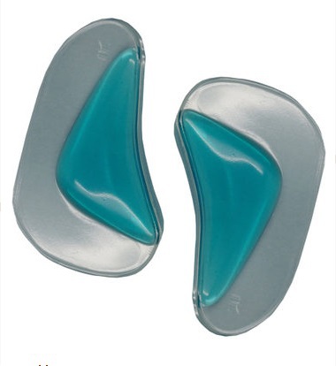
Causes
Several risk factors are associated with PTT dysfunction, including high blood pressure, obesity, diabetes, previous ankle surgery or trauma and exposure to steroids. A person who suspects that they are suffering from PTT dysfunction should seek medical attention earlier rather than later. It is much easier to treat early and avoid a collapsed arch than it is to repair one. When the pain first happens and there is no significant flatfoot deformity, initial treatments include rest, oral anti-inflammatory medications and, depending on the severity, a special boot or brace.
Symptoms
At first you may notice pain and swelling along the medial (big toe) side of the foot. This is where the posterior tibialis tendon travels from the back of the leg under the medial ankle bone to the foot. As the condition gets worse, tendon failure occurs and the pain gets worse. Some patients experience pain along the lateral (outside) edge of the foot, too. You may find that your feet hurt at the end of the day or after long periods of standing. Some people with this condition have trouble rising up on their toes. They may be unable to participate fully in sports or other recreational activities.
Diagnosis
In the early stages of dysfunction of the posterior tibial tendon, most of the discomfort is located medially along the course of the tendon and the patient reports fatigue and aching on the plantar-medial aspect of the foot and ankle. Swelling is common if the dysfunction is associated with tenosynovitis. As dysfunction of the tendon progresses, maximum pain occurs laterally in the sinus tarsi because of impingement of the fibula against the calcaneus. With increasing deformity, patients report that the shape of the foot changes and that it becomes increasingly difficult to wear shoes. Many patients no longer report pain in the medial part of the foot and ankle after a complete rupture of the posterior tibial tendon has occurred; instead, the pain is located laterally. If a fixed deformity has not occurred, the patient may report that standing or walking with the hindfoot slightly inverted alleviates the lateral impingement and relieves the pain in the lateral part of the foot.
Non surgical Treatment
Treating PTTD is almost always easier the earlier you catch it. So, the first step in treatment is to see your doctor as soon as you begin experiencing painful symptoms. However, once your condition has been diagnosed, your podiatrist will likely try to give the upset tendon a bit of a break so it?ll calm down and stop being so painful. This can often be accomplished by immobilizing the foot using tape and padding, braces, or casts, depending on what your podiatrist believes will work best for you, and depending on the severity of your condition. You may also be instructed to reduce inflammation by applying ice to the area (usually 40 minutes on and 20 minutes off, with a thin towel between you and the ice). Or, you might take anti-inflammatory medications like ibuprofen (steroidal anti-inflammatory meds are actually likely to make this problem worse, and are not usually recommended in treating PTTD), or use ultrasound therapy. Once the inflammation has gone down a bit, your podiatrist may recommend using orthotics (prescription shoe inserts) to support your damaged arch. Ankle braces can also be helpful. 
Surgical Treatment
In cases of PTTD that have progressed substantially or have failed to improve with non-surgical treatment, surgery may be required. For some advanced cases, surgery may be the only option. Surgical treatment may include repairing the tendon, tendon transfers, realigning the bones of the foot, joint fusions, or both. Dr. Piccarelli will determine the best approach for your specific case. A variety of surgical techniques is available to correct flexible flatfoot. Your case may require one procedure or a combination of procedures. All of these surgical techniques are aimed at relieving the symptoms and improving foot function. Among these procedures are tendon transfers or tendon lengthening procedures, realignment of one or more bones, or insertion of implant devices. Whether you have flexible flatfoot or PTTD, to select the procedure or combination of procedures for your particular case, Dr. Piccarelli will take into consideration the extent of your deformity based on the x-ray findings, your age, your activity level, and other factors. The length of the recovery period will vary, depending on the procedure or procedures performed.